Introduction to MRI Scanning - Uevolve Radiology
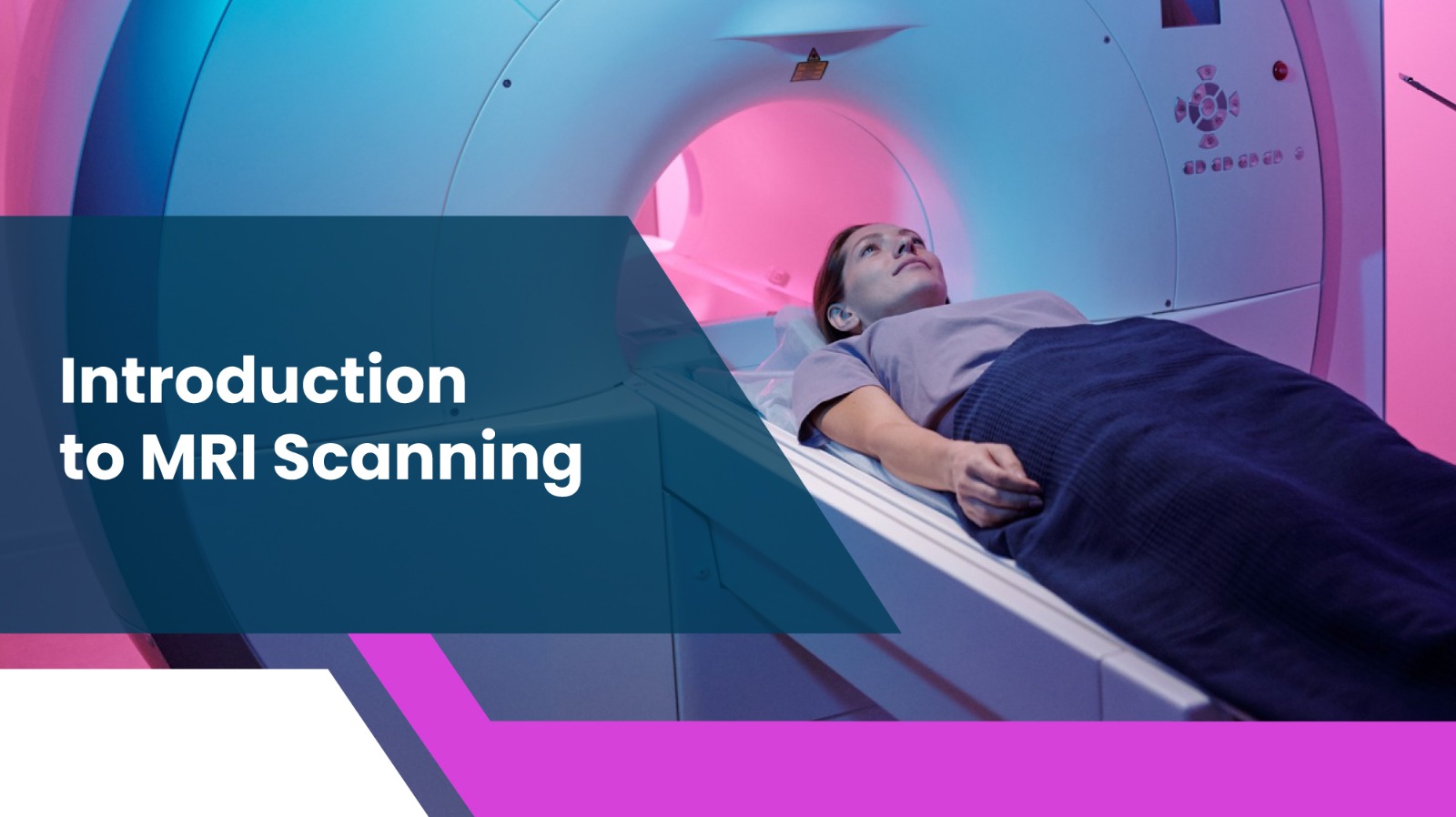
Magnetic Resonance Imaging (MRI) is a medical imaging technique that uses strong magnetic fields and radio waves to create detailed images of the organs and tissues inside the body. Unlike X-rays, MRI scans do not use ionising radiation, making them a safer choice for many patients. MRI is often used to assess soft tissues, such as the brain, spinal cord, muscles, and organs, providing clear images that help doctors diagnose a wide range of conditions.
How MRI Scans Work
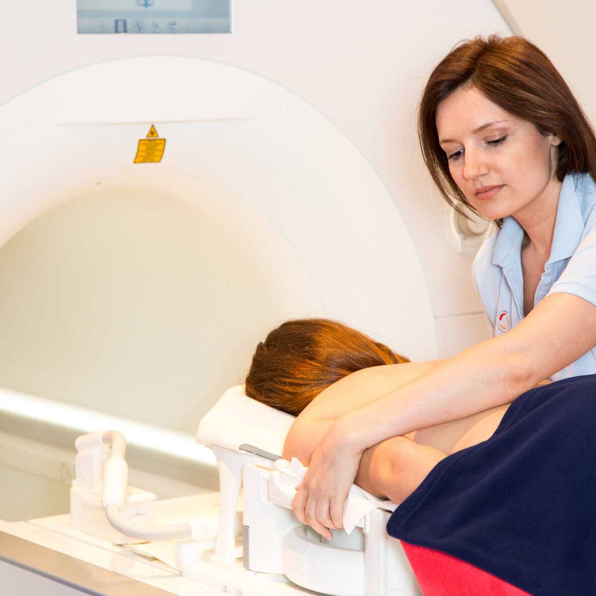
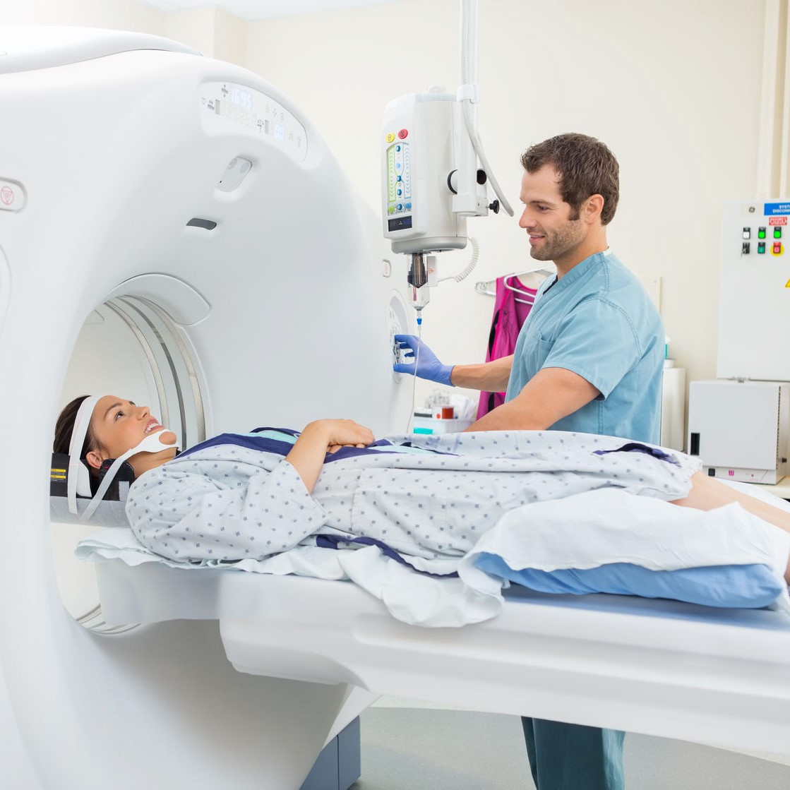
An MRI scanner consists of a large magnet and a computer that creates images by measuring how the hydrogen atoms in the body respond to the magnetic field. When a patient enters the MRI machine, the magnetic field aligns the hydrogen atoms in the body. Radio waves are then sent through the body, causing these atoms to emit signals. These signals are captured by the machine and converted into detailed cross-sectional images, allowing radiologists to examine the structures within the body.
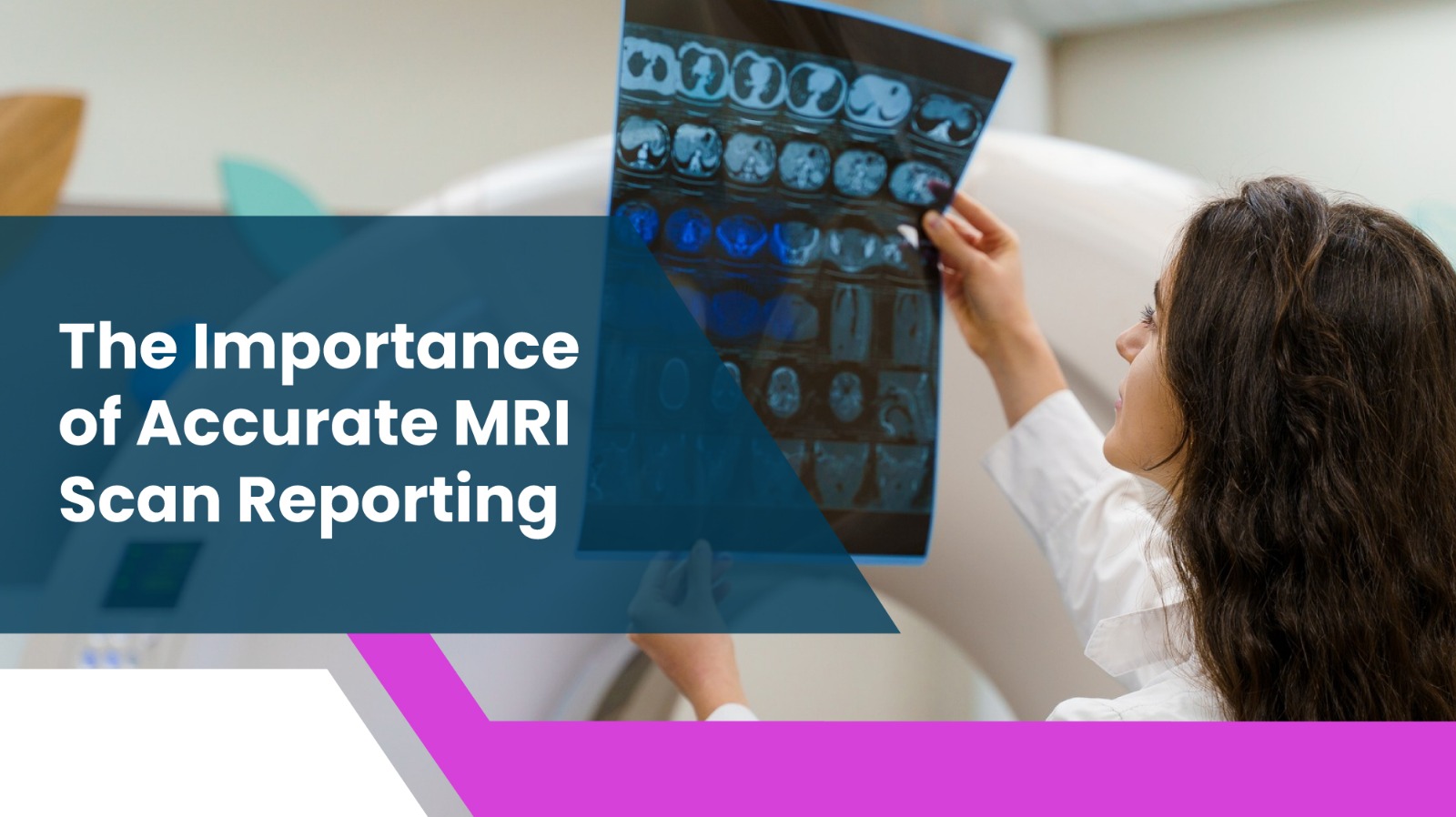
The Importance of Accurate MRI Scan Reporting
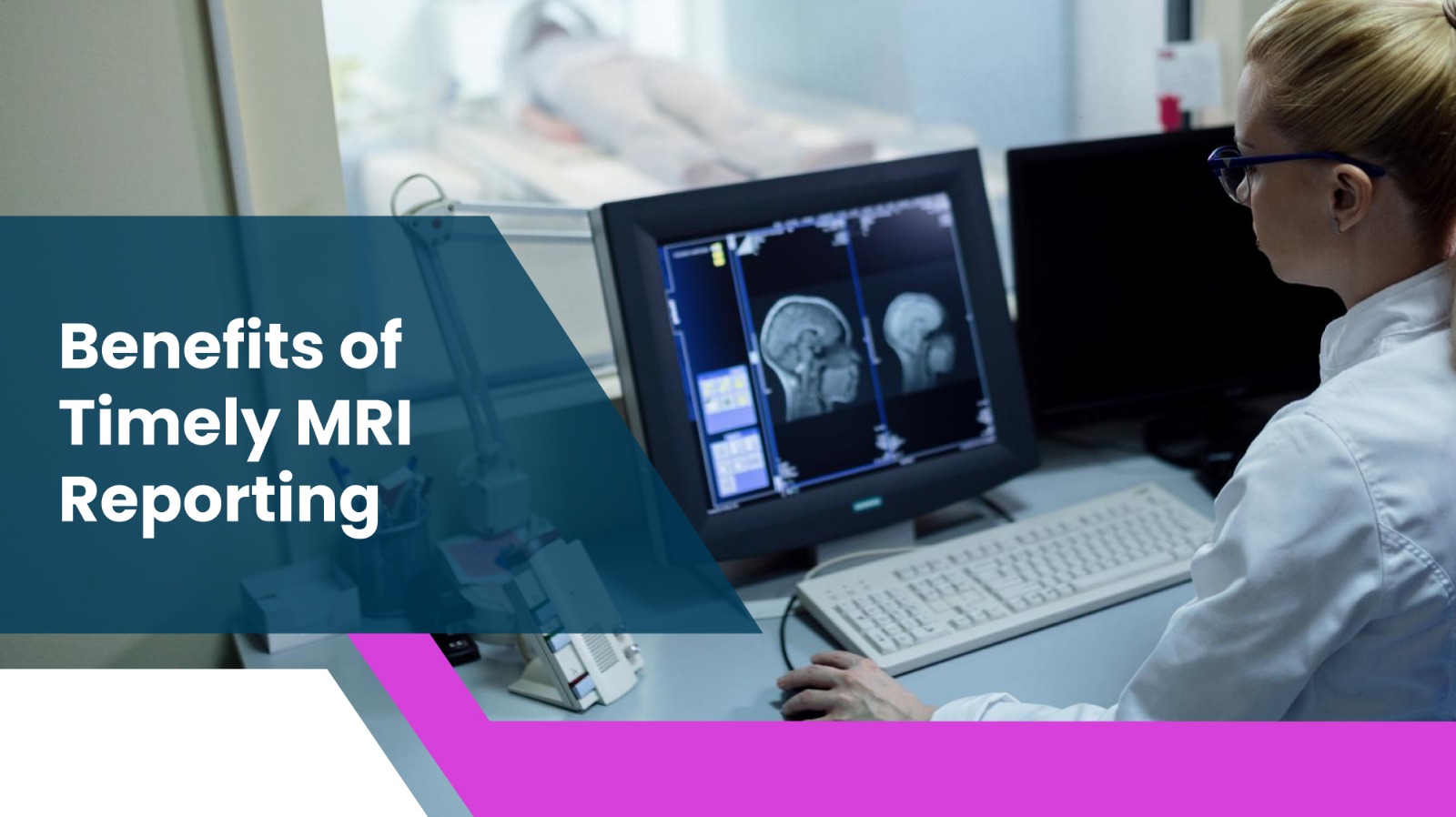
Understanding the Role of a Radiologist
A radiologist plays a crucial role in interpreting MRI scans. These medical professionals are highly trained to read the images and identify any abnormalities or conditions. A radiologist will carefully analyse the MRI scan and create a detailed report that outlines their findings, which can be shared with the referring physician. The accuracy of the MRI report is essential, as it forms the basis for the doctor’s diagnosis and treatment plan.
How MRI Reports Are Created
MRI reports are created by radiologists who interpret the images produced during the scan. The radiologist carefully examines the MRI results for any signs of disease, injury, or abnormalities. The report typically includes detailed descriptions of any detected conditions, along with possible diagnoses. Additionally, the radiologist will highlight any areas of concern and recommend further tests or treatments if necessary. Once the report is completed, it is sent to the patient’s referring doctor for further action.
Common Conditions Diagnosed via MRI Scans

Neurological Conditions (e.g., Brain Tumours, Multiple Sclerosis)
MRI scans are frequently used to diagnose neurological conditions. For example, they are essential in detecting brain tumours, strokes, and multiple sclerosis (MS). An MRI can identify abnormal growths or changes in brain structure, which is vital for timely intervention. Additionally, MRI scans are helpful in diagnosing spinal cord issues, such as herniated discs or spinal stenosis.
Musculoskeletal Issues (e.g., Joint Injuries, Spinal Problems)
MRI scans are invaluable tools for diagnosing musculoskeletal conditions. They can reveal soft tissue injuries such as torn ligaments, tendons, or cartilage, as well as issues in bones and joints. Common examples include diagnosing torn menisci in the knee, rotator cuff tears in the shoulder, or herniated discs in the spine. The clarity of MRI images allows doctors to assess the severity of the injury and determine the best course of treatment.
Cardiac and Vascular Conditions
MRI scans can also be used to evaluate the heart and blood vessels. A cardiac MRI can help identify heart disease, such as coronary artery disease, heart failure, or congenital heart defects. Additionally, vascular MRI is used to assess conditions like aneurysms, blood clots, or abnormal blood flow, allowing doctors to plan effective treatments.
How to Read an MRI Report
Key Terminologies Explained
MRI reports often contain technical terms that may be difficult for the average person to understand. Some common terms include:
T1- and T2-weighted images: Different types of images that show varying levels of contrast in tissues, helping to identify abnormalities.
Contrast agents: Sometimes, a contrast dye is used to enhance the clarity of certain structures in the body, making them more visible on the MRI scan.
Lesions: Refers to any abnormal tissue, such as tumours or areas of inflammation.
What to Expect from Your MRI Report
Your MRI report will typically include a description of the findings, including any abnormalities, their location, and possible causes. The radiologist may suggest additional tests or treatments based on the findings. The report may also include an assessment of the severity of any conditions, helping your doctor determine the next steps in your care.
Benefits of Timely MRI Reporting
Early Diagnosis and Treatment
One of the key advantages of having an MRI scan is the ability to diagnose conditions early. Timely MRI reporting can significantly impact the treatment process, enabling doctors to start treatment as soon as possible. Early intervention often leads to better outcomes, especially for conditions like cancer, neurological disorders, and musculoskeletal injuries.
How Quick Reporting Can Help with Emergency Care
In emergency situations, fast MRI reporting can be a lifesaver. For example, in cases of traumatic brain injury or stroke, rapid MRI scans and reporting allow treating doctors to assess the situation quickly and take necessary action, reducing the risk of long-term damage. Access to fast and accurate MRI reports can significantly improve the chances of a positive outcome for patients.
Why Choose Professional Radiology Services?
Advantages of Consulting Experts in MRI Reporting
Choosing a reliable and experienced radiology service is essential for obtaining accurate MRI results. At Uevolve Radiology, we provide expert MRI reporting with a commitment to precision and clarity. Our team of highly trained radiologists ensures that every MRI scan is analysed thoroughly, and the resulting report is comprehensive and actionable.
Trustworthy and Accurate Reporting at Uevolve Radiology
At Uevolve Radiology, we understand the importance of timely and accurate MRI reporting in ensuring the best care for our patients. State-of-the-art technology and years of experience equip our radiologists to provide detailed, reliable reports that guide your healthcare decisions. You can trust Uevolve Radiology for all your MRI scanning and reporting needs.
For more information, visit Uevolve Radiology Services.

Leave a Comment